Functional Imaging and Instrumentation Group
FIIG Research
Research activities in the field of detector R&D
The "Functional Imaging and Instrumentation" group of the Department of Physics and INFN Pisa is from years working on the
development of new tools for molecular imaging, with focus on the pre-clinical applications of Positron Emission Tomography
(PET) and its combination with the X-ray Computed Tomography (PET / CT), and applications preclinical and clinical imaging
and spectroscopy in nuclear magnetic resonance imaging (MRI and MRS).
Our laboratory serves as an incubator for technologies of molecular imaging. The detectors are designed, developed and,
when the technology is mature enough, are integrated into tools for specific applications. The development of data
acquisition systems and software acquisition and image reconstruction is an integral part of the process of prototyping the
imaging systems.
Among the research and development undertaken by our group, one is the development and application of a new medical imaging
photodetector, the Silicon Photomultipliers (SiPM). The SiPM is a device in silicon consists of a matrix of small avalanche
photodiodes (microcells) operated in Geiger mode. The micro-cells are connected in parallel so that the signal provided by
the SiPM is the sum of the signals of the microcells (see figure 1).
The high gain (about 10^6) attainable at low supply voltages (order of 50 V), the compactness and versatility in design
make these devices particularly suitable for their use in medical imaging systems such as PET and SPECT scanners, probes
Intraoperative probes and, due to their insensitivity to magnetic fields, even in modern hybrid PET/MR systems. Since 2006,
the group led the project INFN DASIPM which aimed to develop and apply the SiPM in various fields of research, spanning
from astrophysics, calorimetry for high energy physics to medical imaging. As part of this project it was produced the
first Italian SiPM in collaboration with the research institute FBK-irst that at present is among the world's leading
manufacturers of this device. Appropriately tailored versions of these detectors will be used in a PET/MR scanner dedicated
to investigations of psychiatric diseases (EU FP7 project TRIMAGE) and a system for verifying the quality of cancer
treatments in hadrontherapy (Ministry project PRIN INSIDE) to be installed at the National Center of Oncology Hadrontherapy
(CNAO) in Pavia (see figure 2).
Working in close collaboration with our colleagues at the Institute of Clinical Physiology (IFC-CNR Pisa), where physicists,
biologists, radiochemical created the "Micro Imaging Lab" laboratory dedicated to pre-clinical investigations, we are now
taking our technology from our laboratory in real systems ready to be used in routine practice.

Figure 1. Sketch of the Silicon photomultiplier.

Figure 1. Silicon photomultiplier matrix.
Research activities in the field of software development
Several areas of Medical Physics require the development of dedicated software. In particular, we are involved in the automatic analysis of diagnostic digital images, eg Pulmonary CT (a), magnetic resonance images (MRI) of the brain (b) and of the muscoloskeletal system (c), in order to create software tools that provide useful information for the quantitative analysis and interpretation of the images.

a) GUI of the lung CAD web service (left) and visualization of CAD findings with a dedicated plugin of OsiriX DICOM viewer (right).

b) Examples of discrimination maps overlaid to a representative structural MR image, obtained from SVM-RFE analysis.
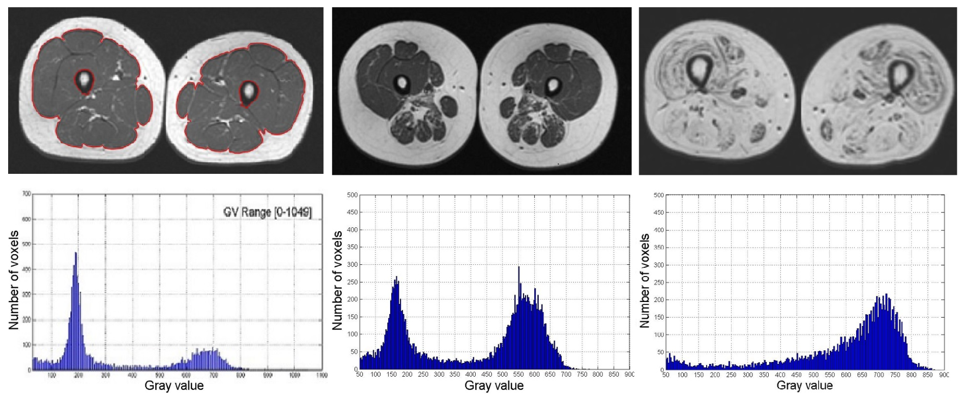
c) Automatic muscle segmentation and intensity hystograms for healthy subject (left) and NMD patients with mild (middle) and severe (right) muscle involvment.
Research activities in the field of molecular imaging instrumentation
According to the most widely accepted definition by S. S. Gambhir: "Molecular Imaging is the defined as the visual
representation, characterization, and quantification of biological processes at the cellular and subcellular levels
within intact living organisms. It is a novel multidisciplinary field, in which the images produced reflect cellular
and molecular pathways and in vivo mechanisms of disease present within the context of physiologically authentic
environments. The term "molecular imaging" implies the convergence of multiple image capture techniques, basic
cell/molecular biology, chemistry, medicine, pharmacology, medical physics, biomathematics, and bioinformatics into
a new imaging paradigm".
With all this in mind we are working on the development of new imaging instrumentation for molecular imaging
with a stronger focus on pre-clinical applications of dual modality Positron Emission Tomography and X-ray
Computed Tomography (PET/CT).
o this aim, our laboratory serves as an incubator for Molecular Imaging technologies. The detectors are
designed, developed and, when mature enough, are implemented in application-specific instruments. Data
acquisition systems, acquisition and image reconstruction software are synergically developed and integrated.
Working in strict collaboration with our colleagues at the Institute of Clinical Physiology
(CNR-IFC, Pisa) where Physicists, Biologists,
Radiochemists have created "Micro Imaging Lab" dedicated to pre-clinical investigation, we are translating our
technology from our laboratory into "real" systems ready to be used in the routine practice.
In last ten years we have developed and turned into prototypes various imaging systems:
PET")
The YAP-(S)PET and example of PET imaging on mice.

The IRIS PET/CT and example of hybrid PET/CT imaging on mice.

The XaltHR and example of CT imaging on mice.
Laboratory of Medical Physics and MR Technologies
The research activity of the Laboratory of Medical Physics and MR technologies of IRCCS Stella Maris and IMAGO7
research foundation has been developing around two Magnetic Resonance (MR) scanners. The first is a clinical scanner
at 1.5 Tesla (Signa Horizon LX General Electric HealthCare), optimized for neurological applications and operative at IRCCS
Stella Maris from 1996. Since two years ago, an Ultra-High Field MR (UHF_MR) scanner at 7 Tesla for human application
(Signa7T MR950 General Electric HealthCare) is active at IMAGO7. It is committed only for research purposes, being the
first UHF-MR system in Italy.
The long experience on clinical MR scanners allowed to execute and optimize: i) neurometabolic exams by Magnetic
Resonance Spectroscopy (MRS) of hydrogen (1H) and phosphorous (31P); ii) functional imaging (fMRI); iii) metabolism
studies as the perfusion weighted imaging by the measurement of the Cerebral Blood Flow (CBF) and iv) the study of
cerebral microstructure by using diffusion techniques such as the Diffusion Tensor Imaging (DTI) and the Fiber Tracking.
Principal focus of research activity is the application of all these MR methods to the field of neuroscience and in
the neurological and psychiatric disease of developmental age.
Thanks to the use of UHF-MR, many and new potentialities are opened in terms of increment of Signal to Noise Ratio
(SNR), increment of spatial, temporal and spectral resolutions, as well as in the exploitation of new contrast mechanisms,
able to depict the brain structure at sub-millimeter level, such as the techniques of Susceptibility Weighted Imaging,
(SWI). However, at UHF-MR, there are also many technical challenges in the application of standard sequences,both in
terms of safety (as the monitoring and the control of energy deposition on the patient) and in terms of distortions
of the images and in-homogeneities of the signal. Furthermore, to take full advantage from UHF-MR, pulse sequences
have to be optimized and hardware components such as dedicated and new designed RF coils have to be developed for
specific MR applications. Thus, a specific R&D activity in collaboration with INFN and GE HealthCare is on-going.
For more informations:

Iper-defined brain image acquired by the 7 Tesla MR Scanner.
Research activities in the field of monitoring for particle therapy treatment
A promising technique for cancer treatment is using charged particles like protons and carbon ions, rather than photons,
and several facilities operate in this field since many years (more information at: http://www.ptcog.ch). This growing
interest in charged particle therapy is based on the highly improved dose conformation to the target volume potentially
resulting in an improved sparing of organs-at-risk. Treatments with charged particles are, however, much more sensitive
to uncertainties than photon treatments: an active research area in particle therapy is therefore how to monitor the dose
delivery. Positron Emission Tomography (PET) is a non-invasive way of in-vivo verification of the dose delivered to the
patient. During ion beam irradiation, various Beta+ emitting isotopes (15O, 11C, 13N etc) are generated in the patient.
Although dose and activity are indirectly related, it is possible to obtain useful information about the proton range and
delivered dose from beta+ activity measurements. By comparing the measured PET activity distributions with a pre-calculated
Monte Carlo distribution, it is possible to check whether the delivered dose differs from the planned dose, i.e. to perform
quality assurance of the treatment.
In this context, in Pisa (University of Pisa and INFN: National Institute of Nuclear Physics), a group of researchers has
developed two different systems for this purpose: DoPET and INSIDE.
DoPET is a compact planar PET system, that can be installed in the beam delivery system, and was developed in the framework
of the project RDH (Research and Development in Hadrontherapy) funded by INFN-Italy. This system is capable of acquiring data
during the treatment as was demonstrated for proton irradiation with the CATANA cyclotron and with the CNAO synchrotron.
DoPET, consisted of two planar 16 x 16 cm2 detector heads each composed of nine independent modules. Each module consisted
of an H8500 PMT coupled to a 23x23 LYSO crystal matrix (2 mm pitch) with the readout performed by custom developed
electronics. The Data Acquisition was designed to keep the acquisition dead time low and to have a relatively narrow
coincidence window (few ns). DoPET has a sensitivity and spatial resolution so as to reconstruct the Bragg peak position
to 1 mm in less than few minutes after the dose delivery for proton irradiations, limiting the biological washout problems.
INSIDE combine an in-beam PET scanner and a particle tracking system in a unique imaging device to measure on-line the
particle range with high precision. This bi-modal monitoring system is being developed in the framework of the project
INSIDE (Innovative Solutions For Dosimetry In Hadrontherapy) funded by MIUR, the Italian Minister for University and
Research) under the PRIN program (Research Projects of National Relevance). The PET scanner, an evolution of the DoPET
system, is tailored for the imaging of head and neck tumours. It is composed of two planar detectors, 10 cm wide
(transaxially) x 25 cm long (axially, along the beam direction). The PET heads are composed of 2x5 detection units, each
made of arrays of 256 LSF pixel crystals, 3x3x20 mm3 each, coupled one to one to analog solid state photodetectors called
Silicon PhotoMultipliers (SiPM). A custom designed electronics system is being developed to cope with the prompt particle
rates typical of a therapeutic beam (~10 MHz/cm2).
The PET activity map created in the target is complemented by the beam profile, obtained by tracking the secondary particles
(protons) coming from the interaction of the primary beam with the target nuclei and from projectile fragmentation in case
of carbon beams. The tracking system is composed of 6 planes of orthogonal squared scintillating fibers (each plane has 384
fibers) coupled to 1 mm2 SiPMs. The sensitive area is 19.2 x 19,2 cm2. An electromagnetic calorimeter made of LFS crystal
arrays coupled to Position Sensitive PMTs is placed downstream the tracker and provides the residual energy of the charged
particles.
The INSIDE system is designed to fit in the treatment room of the Centro Nazionale di Adroterapia Oncologica (CNAO, Pavia,
Italy), at the moment the only dual ion (protons and Carbons) PT therapy facility in operation in Italy.


Left: DoPET during a data taking at Protontherapy Centre in Trento. Right: DoPET prototype as a whole.